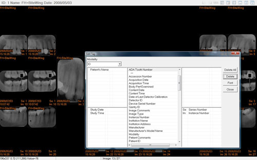
 For the uninitiated, DICOM stands for Digital Imaging and Communications in Medicine. This digital format provides standardization of x-ray images for exchange and provides a means of documenting a considerable amount of embedded information within an individual film or imaging series (e.g. CBCT). AS H. Oosterwijk, A Farman, and P Gihring state in the International Congress Series article titled ‘Standardization of image exchange for dentistry using DICOM (Vol. 1268, June 2004 (p1174-1178)), “in addition, use cases are defined to aid in defining integration profiles for Dentistry”.
It took 23 years for the medical establishment to develop and present DICOM and the format continues to be refined with advances in digital imaging systems. And essentially since 2004, the DICOM standard has also been accepted as a standard for dental x-ray applications. As a result, most dental imaging acquisition modalities allow for DICOM image identification and there are a number of PACS (Picture Archiving and Communication Systems) that have been released to allow for the storage, retrieval, and viewing, among other things, of these DICOM images.
As Dale Miles points out in his article in ‘learndigital.net’ titled DICOM in Dentistry: Interoperability and X-rays (2004), the reason Dentists should care about this development is that now communication between dentists and their medical colleagues can occur with full encryption that is HIPPA compliant. Dentists, as he states, can now have an “almost immediate second opinion” with internet transmission of x-ray imaging because with this security, the x-rays can be transmitted with only remote possibility of hacking.
In February the DICOM WG-22 group will meet again to discuss issues relating to the DICOM standard in Dentistry. In advance of this, Dr Allan Farman has released the following information as an update.
DICOM WG-22 (Dentistry) Strategic Plan:
Co-Chair (User): Allan G. Farman, BDS, PhD (odont), DSc (odont), Univ. Louisville
[email protected]
Co-Chair (Vendor): Xavier Carayol: Carestream/Trophy
[email protected]
Secretariat: American Dental Association
Date of Last Update: October 12, 2024
Scope:
To address all issues relating to the DICOM Standard for dental and maxillofacial applications including imaging and use of imaging in diagnosis, treatment simulation, treatment guidance, and tissue restoration using CAD/CAM. These include (but are not limited to) ordering, acquisition, processing, storage, communication, display and reporting.
Roadmap:
The implementation of dental and maxillofacial relevant objects (e.g. DX, IO, SC, SR) will be emphasized for dental care environments. Specifications within DICOM are needed to promote image interoperability between digital imaging systems for dentistry and throughout the processes of treatment simulation, treatment guidance and the CAD/CAM prosthetic chain. Coordinated education and demonstration projects for vendors and users are essential to achieve broad adoption of the DICOM Standard in Dentistry. Extensions/refinements to existing objects will be introduced to accommodate existing and emerging digital imaging and related techniques used in Dentistry.
Current Work Items:
Support of WG 24 initiatives on DICOM for implants (dental use cases).
Imminent Goals:
• Introduction of a DICOM Supplement to support the full CAD/CAM prosthetic chain. DICOM surgical workflow issues in virtual and solid anatomic model fabrication
• Refine Structured Display and develop a Dental Query-Retrieve application
• Support of joint WG 24/WG 22 initiative on optical surface scanning
Future Goals:
• Development of guidelines for standardization of digital photographic structured displays for intra-oral and extra-oral projections. DICOM STRATEGIC DOCUMENT PAGE 28
• Applications of WADO for dentistry.
• Creation of templates for structured reporting.
• Development of guidelines for presentation states including overlays used in dentistry.
• DICOM surgical workflow issues in dental implantology
• Extending Supplement 116 (3D X-ray) into specific dental applications.
• Evaluation of existing objects or current work items for use in dental imaging.
Risks:
Implementation of existing standardized DICOM objects is just beginning in Dentistry. While there is heightened awareness of DICOM in the vendor and user communities, new vendors are entering the marketplace with increasing frequency. There is a need to introduce the new vendors to the concept of DICOM and also to explain both the advantages and limitations of DICOM to users. Further, it is necessary to continually restate that interoperability holds more advantages than disadvantages as part of the business model and also for the ethical use of radiation in diagnostic situations.
Relationships to Other Standards:
WG-22 anticipates working closely with the American Dental Association Standards Committee on Dental Informatics initiatives for interoperability of all electronic media, inclusive (but not exclusive) to diagnostic images. DICOM WG 22 and has some overlap in membership with ADA SCDI WG 12.1 (Digital Imaging), and also has task groups within the Japanese Society of Oral and Maxillofacial Radiology, the American Academy of Oral and Maxillofacial Radiology, the American Association of Orthodontists and industry in Europe. It is recognized that the goal of interoperability for Dentistry will involve concepts that transcend DICOM and include HL7 and IHE
Provided by: Allan G. Farman, BDS, PhD, MBA, DSc, Diplomate ABOMR
Prof. Radiology & Imaging Science
Univ. Louisville School of Dentistry: SUHD
Editor's note: Dr Brent Dove provides additional useful commentary on DICOM at: http://ddsdx.uthscsa.edu/DICOM.html


