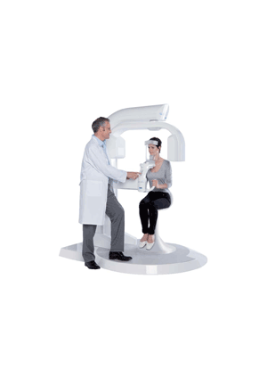
3D diagnostics require confidence and reliability, but also precision and simplicity. With the new WhiteFox from SATELEC (ACTEON Group), oral surgeons, implantologists, orthodontists and ENTs have a digital cone beam computed tomography of the latest generation that gives them precise and reliable 3D x-ray data from the entire oral-maxillofacial area in the shortest time with one scan.
In endodontics, functional diagnostics, oral surgery, implantology and orthodontics, the powerful and elegant CBCT multifunctional device ensures reliable diagnoses in all areas of dentistry with five field of view sizes (FoV) .
for definitive, predictable therapeutic and surgical results. Since the Acteon Group acquired the Italian high tech company de Götzen® four years ago, the company, which had already enjoyed great success with its X-Mind AC/DC intraoral x-ray systems, has concentrated on developing more innovative x-ray systems.
With WhiteFox, Satelec now offers a digital volume tomography (DVT) that enables scans in five field of view sizes (from 60 x 60 mm for half arch to 200 x 170 mm for cephalometric images) to provide the best image quality at the lowest possible radiation dose for the patient.
Maximum image quality - minimum radiation dose WhiteFox is the first Cone Beam Computed Tomography (CBCT) system to use the Hounsfield scale, long-established in medical computer tomography. The Houndsfiled scale value enables a very precise and constant measurement of the tissue density in grayscale and allow comparison of pre- and post-surgical analyses with one another.
The practitioner can better decide from the differentiated projection of the bone quality whether immediate implantation is a promising option for the patient.
In addition, the practitioner also has a clear segmentation of soft and hard tissue for a better diagnosis of the jaw and an exact projection of the airways based on virtual slices (virtual endoscopy). Other possible indications include: gnathological and plastic surgery with additional soft tissue filters, comparative analysis of the condylar heads, projection of all sinuses and the middle and inner ear as well as volume measurement of the biomaterials for the sinus lift procedure.
The large 200 x 170 mm field of view enables WhiteFox to produce a precise two-dimensional x-ray image for cephalometric analysis at a 1:1 ratio - without distortion, magnification or stitching - in just one scan!
All-inclusive: Software upgrades and patient comfort The selectable FoV's pulsed mode acquisition, the special resolution setting and the short scan time of only a few seconds mean that the patient receives the minimum radiation exposure.
The perfect combination of elegant form and high functionality in the open arch design means that the patient can simply sit down comfortably and the risk of blurry images is considerably lessened. Digital system expertise from a single source: Unlike other DVT devices, both the primary reconstruction FDK algorithm and the visualization software for WhiteFox were developed within the company so the many software tools are carefully designed to work together, minimizing readout and transmission errors. Unlimited software updates can be installed at any time during the immediate remote maintenance of the working computer.
At the same time, the user has four additional licenses to install on other practice computers, paired with first-class customer service and support from SATELEC. Intuitive image processing, comprehensive functionality Another impressive feature of the new DVT stand unit is the fast reconstruction time. After less than a minute, the result is visible on the screen.
The WhiteFox imaging software then makes the analysis easier with the help of various powerful image processing functions for visualization, diagnosis and treatment planning.


