Guided surgery has been around for a long time. However, very few dentists in the UK place implants using surgical guides. The reasons for this are multiple, ranging from dentists not wanting to follow the procedure, or not having confidence in the procedure, the increased costs of guide fabrication, and the time delay and extra appointments needed to obtain a fully functional and reliable surgical guide.
In this case report, I shall demonstrate a surgical guide manufactured in-house using the CEREC Bluecam (Sirona). These guides do not require any impressions to be sent to a third party and can be made rather cheaply in the surgery within around 30 minutes. The guide can then be used in conjunction with specific drill keys, which are compatible with the guided surgery drill sets from all leading implant manufacturers.
In this particular case, Facilitate (Astra Tech/DENTSPLY Implants) was used to place the implant. Once the implant was osseointegrated, the final restoration was fabricated chairside using the CEREC MC XL milling machine (Sirona) and an IPS e.max CAD block (Ivoclar Vivadent).
Case report
A young female patient had lost tooth 36 a few years ago and wanted an implant solution. Her medical history was clear and she had a mildly restored dentition with no current dental pathology. Her BPE scores were low, with excellent oral hygiene.
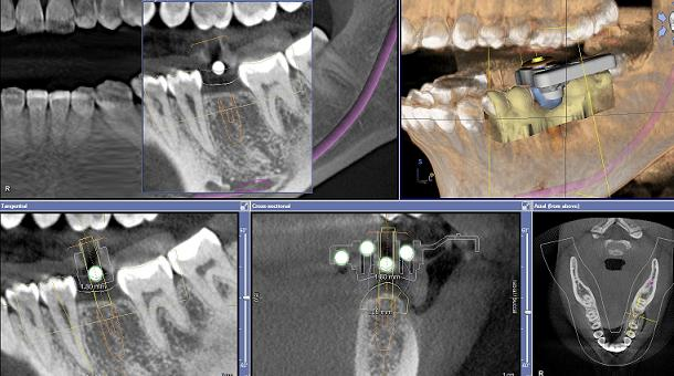
The patient was scanned using the CEREC Bluecam and a proposal for the missing tooth was created. A collimated CBCT scan of the lower jaw was taken using GALILEOS (Sirona) with a CEREC Guide reference body set in thermoplastic over the edentulous area.
The reference body is identified by the software and a virtual implant placement along with the CEREC crown proposal is imported into the software. This allows the clinician to place the implant virtually, with reference to the ideal final crown position. In this case, it was deemed that a screw-retained restoration would be desirable; hence, the screw-access hole was positioned through the centre of the crown.
Once the implant position had been decided, the information was ported to the CEREC software and using a CEREC Guide Bloc a drill body was milled by the CEREC MC XL milling machine. Once this has been milled, it will lock tightly into the thermoplastic drilling template. At this point, the surgical guide is complete and can be used on the patient.
In this particular case, an OsseoSpeed TX implant (DENTSPLY Implants) (4.0 × 11 mm) was placed using the surgical guide. The patient was prepared in accordance with a standard sterile protocol and the area anaesthetised as one would for a regular implant placement. The surgical guide snaps firmly over the existing teeth, expanding over- and undercuts, becoming a very stable platform through which to drill. The Facilitate soft-tissue punch was used to remove the overlying soft tissue, and a standard drilling protocol using the Sirona drill keys was followed.
A high primary stability of 40 N cm was obtained and a 4 mm healing abutment was placed immediately. The patient healed with no pain, no swelling and no discomfort. The post-operative long-cone periapical radiograph corresponded well with the preoperative planning with an ideal angulation for a screw-retained crown. After two months of healing, a fixture-level open-tray impression was taken and cast up using an Astra Tech replica. A standard metal abutment was inserted into the replica and cut back by 3 mm from the occlusal table. This was then powdered and scanned using the CEREC Bluecam, and an IPS e.max CAD C 14 block was milled.
The CEREC 4.2 software was instructed to mill a hole that corresponded to the screw-insertion path on the abutment. This was finished using a high-speed diamond bur with copious irrigation. The crown was glazed and sintered, allowed to cool and bonded to the abutment using Variolink II (Ivoclar Vivadent). The final crown was screwed directly onto the implant and a final check for contacts and occlusion was done.
This process shows just how far CAD/CAM technology has come. An implant can be planned, inserted and restored all in-house, using the current available technology. The final result is equal to any laboratory-based restoration, albeit for simple units. The process does have its limits in terms of multiple-span bridges and placement of multiple implants, especially in edentulous areas. As the technology develops, with further advances being made, the scope of what is possible for the implant dentist is always expanding.


