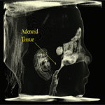
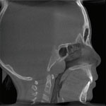
Solving Airway Mystery with i-CAT®Posted in Case Of The Week on April 30, 2024 by m.khodeer Pre-operative picture of patient and CBCT scan showing his severe airway restriction This seven year old male patient was experiencing snoring, mouth breathing, and nighttime bruxism, as well as frequent sinus and ear infections. After numerous doctors were unable to reach a definitive diagnosis, we captured an i-CAT Quick Scan. This 4.8-second scan is approximately half the dose of the i-CAT 8.9-second scan – 74μSv, 2007 tissue weight – and is roughly equivalent to a traditional 2D X-ray series with rectangular collimation, or a pan/ceph/bitewings combination. 1,2 . The scan showed a very narrow trachea and airway with adenoid hyperplasia that caused a significant airway obstruction, which explained several of his symptoms. After viewing the anatomy in three dimensions, a treatment plan in conjunction with an ENT was developed that included adenoidectomy, coblation of turbinates, and orthodontic palatal expansion. Since treatment, the child progressed, with improvements in quality of life, breathing, sleeping, and tasting. Pre- and post-operative scans of airway showing marked improvement and post-op picture of patient Two years later, a follow-up included another 4.8-second Quick Scan. The highly-developed software showed that the airway almost tripled in volume from 8 cc to 23 cc, and the smallest cross sectional area (the bottleneck) went from 23mm2 to 168 mm2 . The obstruction was removed, the palatal shelf, being the floor of the nose, was expanded though Phase I orthodontic therapy, the mandible was unlocked from its transverse discrepancy, and the vector of mandibular growth was improved though nose breathing. The TMJs probably received less stress and grew better as a result of treatment, teeth gained more room for eruption, and his profile and the color of his skin both look better. He is more alert and rested during the day from consistent sleep patterns. Why 3D, Why i-CAT® Dr. Juan-Carlos Quintero DDS, MS |


