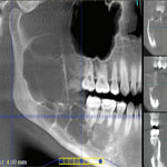
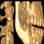
Surgical-Planning Precision Delivered by i-CATPosted in Radiology on April 28, 2024 by m.khodeer Ameloblastoma revealed in i-CAT® scan The patient in this case study was referred to me by her general dentist who discovered an unusual spot on a 2-D x-ray. The patient was asymptomatic. My suspicion, that the spot was an ameloblastoma, an aggressive odontogenic tumor, was confirmed by a biopsy. An i-CAT® scan revealed the very destructive nature of the tumor and the resulting expansion and breakdown of the jaw. All of the data provided by the scan guided me through an innovative treatment plan and gave me the confidence to carry it out to its successful completion. Because of the destruction, a portion of the bone needed to be resected. The scan provided the necessary measurements to calculate for resection. The 3-D images also gave me the tools to create a stereolithic model and prepare the bone plate for easy insertion into the defect at the time of surgery. Reconstruction bar on model, in situ, and in post-op i-CAT® scan – true representation of the anatomy To begin the process, I transferred the 3-D scan information to a CD and sent it to a company that transforms the scan into a 3-D stereolithic model of the patient’s jaws. Then, I simulated the surgical plan on the plastic model, and then used the same model to prepare a metallic temporary replacement jaw and temporomandibular joint. I entered the operating room for the actual surgery, as prepared as possible. I confidently anticipated a successful surgical outcome, knowing that the process would be quicker, easier, and yield no surprises. The actual surgery efficiently and effectively reflected that which I did on the model at my desk in the office. In cases where pathology involves the breakdown of jawbones, visualizing the situation beforehand is a tremendous asset. With a scan that is virtually identical to the patient’s true dental and jaw anatomy, a successful outcome is much more probable. Besides the scans themselves, Cone Beam’s capability for integration with guided surgical techniques and other state-of-the-art applications increases my horizons and my opportunity to add a new dimension to my capacity as a surgeon. Why 3-D, Why i-CAT® Steven A. Guttenberg, DDS, MD |


